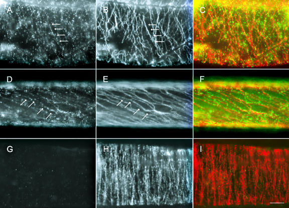
Figure 2.
Immunolocalization of GhKCBP in cotton fibers. Fibers of 10 DPA (A–C and G–I) and 21 DPA (D–F) were stained with anti-GhKCBP (A, D, and G) and anti-α-tubulin (B, E, and H). Pseudocolored images (C, F, and I) showed anti-GhKCBP in green and anti-α-tubulin in red. Note that GhKCBP signal could be easily traced along transverse cortical microtubules at 10 DPA, and along steeply pitched cortical microtubules at 21 DPA (arrows). In control experiments, fibers were stained with depleted anti-GhKCBP (G) and anti-α-tubulin (H). See “Materials and Methods” for methods of depletion. Scale bar = 10 μm.

