Figure 4.
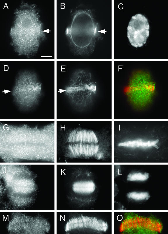
Immunolocalization of GhKCBP in cotton cells undergoing mitotic cell division. Cotton root cells were stained by immunoflurescence techniques for GhKCBP (A, D, G, J, and M) or for α-tubulin (B, E, H, K, and N) or were stained with DAPI to detect DNA (C, I, and L). Pseudocolored images (F and O) show anti-GhKCBP in green and anti-α-tubulin in red. GhKCBP could be detected in the microtubule preprophase band (arrow, A, B, and D–F). Abundant GhKCBP signal was present in metaphase cells (G–I). GhKCBP clearly was present among phragmoplast microtubules (J, K, M, to O). Scale bar = 10 μm.

