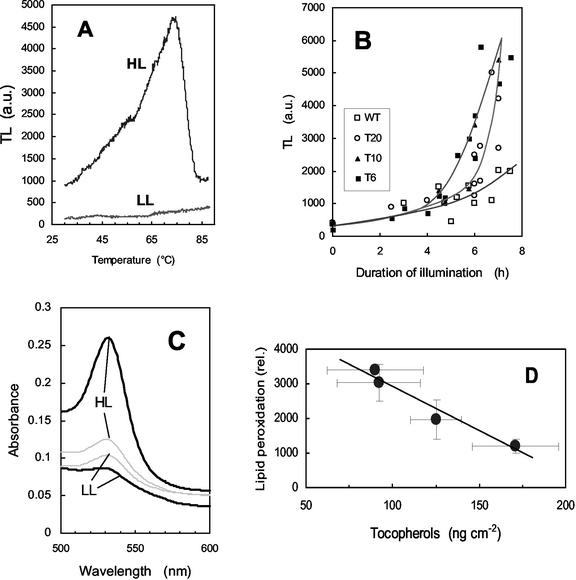
Figure 3.
Photooxidation of tobacco leaf discs exposed to a high PFD of 1,000 μmol m−2 s−1 and a low temperature of 10°C. Leaf discs were taken from plants grown in low light at 25°C. Lipid peroxidation was measured by the amplitude of the 70°C to 80°C thermoluminescence signal as shown in A (HL, T6 leaf discs exposed for 7 h to the light stress; LL, control T6 leaf grown in low light at 25°C). B, Time course of the increase in thermoluminescence (80°C peak) during chilling stress in high light. White squares, WT; black squares, T6; black triangle, T10; white circle, T20. C, Malondialdehyde (MDA) level, as indicated by its light absorption at 532 nm after reaction with TBA in acidic medium, in T6 (thick line) and WT (thin line) leaves before (LL) and after (HL) high-light stress at low temperature. D, Plot of the lipid peroxidation status of tobacco leaves (measured by thermoluminescence after 6 h in high light at low temperature) versus the tocopherol concentration.

