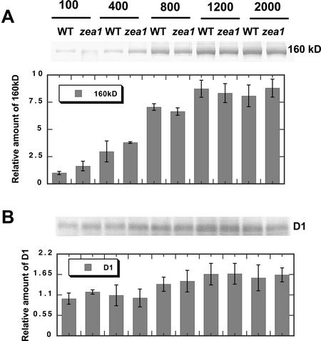
Figure 3.
Quantitative western-blot analysis of thylakoid membrane proteins from D. salina WT and zea1 mutant grown under different light intensities. Proteins were probed with specific polyclonal antibodies against the 160-kD PSII repair intermediate (A) or against the PSII D1/32-kD reaction center protein (B). The corresponding densitometric quantitations of the bands are given as a bar graph on the bottom of each panel. Lanes were loaded on an equal Chl basis (4 nmol Chl lane−1).

