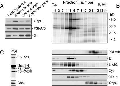
Figure 3.
Association of Ohp2 with PSI. A, Localization of Ohp2 in appressed and nonappressed regions of thylakoid membranes assayed by immunoblotting. As references, the distribution of subunits-A/B from the PSI reaction center (PSI-A/B) and the D1 protein from PSII (D1) reaction center were assayed. B, Location of Ohp2 in PSI. Thylakoid membranes isolated from control leaves grown under ambient light conditions (100 μmol m–2 s–1), were solubilized with n-dodecyl β-d-maltoside, and the released protein complexes were separated by a Suc density gradient centrifugation. The protein composition in collected fractions was analyzed on SDS-gel stained by Coomassie Blue (top). The distribution of PSI, PSII, and its antenna, cytochrome b/f, and ATP synthase complexes were analyzed by immunoblotting using polyclonal antibodies directed against the A and B subunits of PSI reaction center (PSI-A/B), the D1 protein of PSII reaction center, the chlorophyll a/b-binding protein of PSII (Lhcb2), the subunit f of the cytochrome b6/f complex (cyt f), and the α-subunit of the CF1 ATP synthase complex (CF1-α; middle panels). Location of Ohp2 was assayed by immunoblotting (bottom). C, Composition of the fraction 11 tested by antibodies directed against subunits A, B, C, D, E, F, H, and L of PSI (PSI-A/B, PSI-D/F/L, or PSI-C/E/H, respectively), light-harvesting antenna proteins of PSI (Lhca1–4), and Ohp2.

