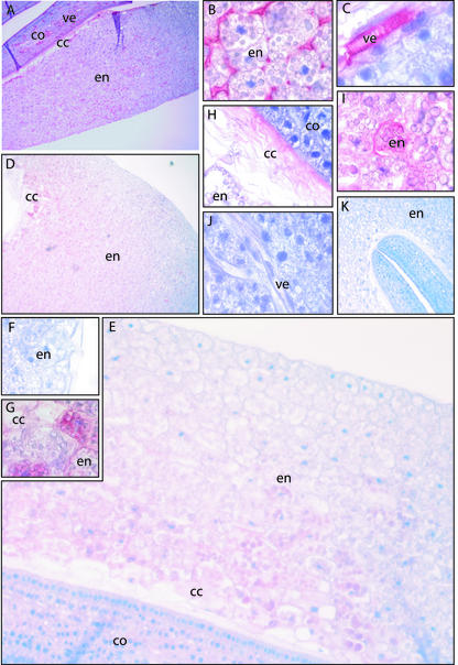
Figure 7.
Immunohistochemistry using antibodies against ElLTP1 and ElLTP2 on sectioned seedlings collected 4 d after sowing. Red color indicates detection of antigen, and blue color is counter staining of nuclei with Mayers hematoxylin. A through C, Result after staining with the anti-ElLTP1 antibody. D through J, Result with the anti-ElLTP2 antibody. K, Negative control, which is stained without primary antibody. A, Overview of anti-ElLTP1 staining in the endosperm (en), collapsed cell region (cc), and cotyledons (co). B, Detection of the ElLTP1 in apoplastic space in the middle part of the endosperm. C, Detection of ElLTP1 in the vessel elements (ve) of the cotyledons. D, Overview of anti-ElLTP2 staining in the endosperm and collapsed cell region. E, A closer view of the detection of ElLTP2 in endosperm, cc region and the cotyledons. F, The weak anti-ElLTP2 staining in the outmost part of the endosperm. G, A strong anti-ElLTP2 signal was obtained from the inner part of the endosperm. H, Detection of ElLTP2 in the cc region. I, In the endosperm, ElLTP2 was detected between and also inside cells. J, ElLTP2 was not detected in vessel elements or other cells of the cotyledons.

