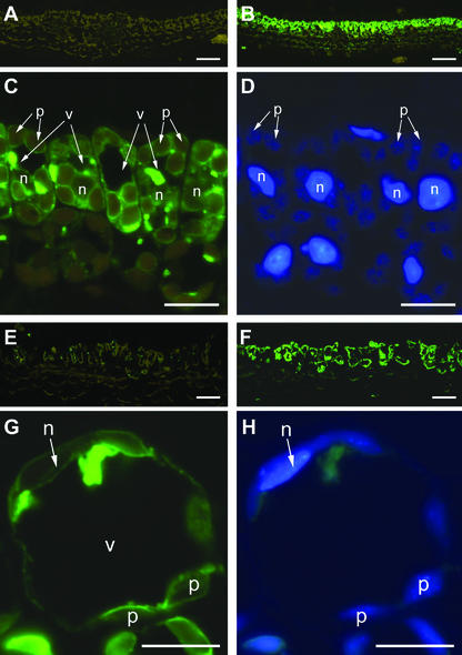
Figure 5.
Immunofluorescence labeling of Gleheda in leaves of ground ivy. Developing leaves (A–D) and fully developed leaves (E–H) were used for embedding. Cross sections of leaves of a no-lectin (clone CA 1; A and E) and a highlectin plant (clone EF 5; B–D and F–H) were probed with anti-Gleheda-antibody, followed by a labeling with fluorescence-labeled secondary antibody. Labeling in the sections of the low-lectin clone exhibits only the yellow-brown autofluorescence of chloroplasts (A and E), whereas the strong green fluorescence label within the palisade parenchyma in the sections of the high-lectin clone (B and F) indicates the location of the lectin. Higher magnifications of B and F are shown in C and G, respectively, to visualize the subcellular location of Gleheda. Note the high amount of fluorescence-labeled Gleheda within the vacuoles (v). DNA-containing organelles (n, nucleus; p, plastids) were identified by concomitant 4,6-diamidino-2-phenylindole (DAPI) staining (shown in D and H). Bars = 50 μm in A, B, E, and F; and 10 μm in C, D, G, and H, respectively.

