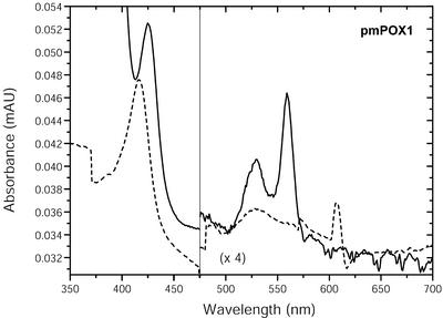
Figure 4.
Absorption spectra of partially purified pmPOX1. Samples (1.1 mg protein mL–1) containing the native enzyme (dashed line) were measured in 50 mm sodium phosphate buffer (pH 7.0) with buffer as reference. Ferric enzymes were reduced by the addition of approximately 1.5 mm dithionite (straight line). The spectra were measured at 50 nm min–1. n = 2 independent preparations showing identical results. The spectra indicate the presence of heme groups as the prosthetic group.

