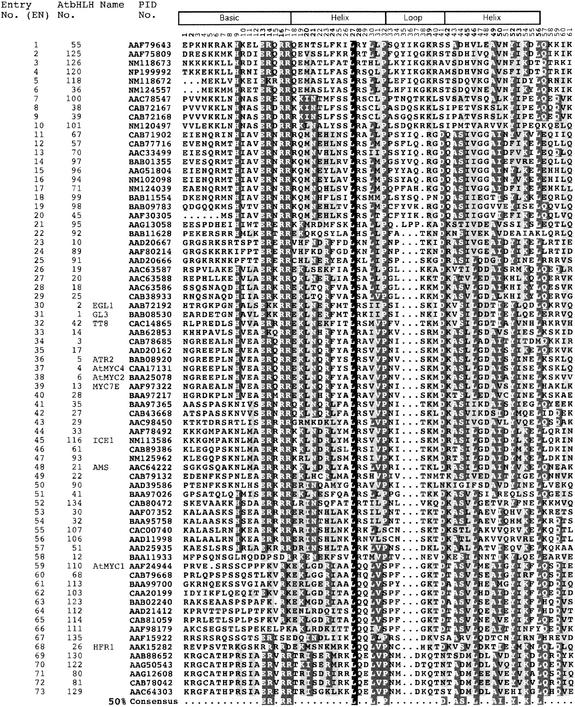
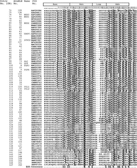
Figure 1.
Multiple Sequence Alignment of the bHLH Domains of the 147 Members of the AtbHLH Protein Family.
Each protein is identified by its PID number and AtbHLH number (Heim et al., 2003). The EN assigned in this study is based on the order in which the proteins are shown in this alignment. The scheme at top depicts the locations and boundaries of the basic, helix, and loop regions within the bHLH domain. The numbers below the scheme (1 to 61) indicate the position within the bHLH motif as defined in this study. For those proteins for which a name has been given, the name is provided after the PID number. The shading of the alignment presents identical residues in black, conserved residues in dark gray, and similar residues in light gray. Dots denote gaps. The Arabidopsis consensus motif at bottom is based on the residues with 50% conservation among the 147 proteins shown.


