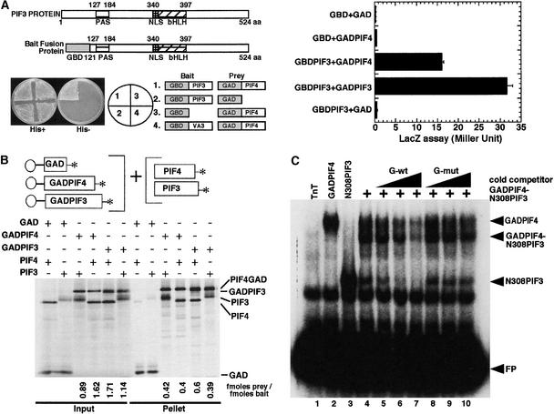
Figure 5.
PIF4 Heterodimerizes with PIF3.
(A) PIF3 and PIF4 interact in a yeast two-hybrid assay. The left panel shows interaction in a plate growth assay. The combination of constructs used in each section is indicated in the circle (middle) and at right. The right panel shows Miller units in a quantitative liquid β-galactosidase assay. GBD and GAD denote GAL4 DNA binding and activation domains, respectively. GAD:PIF4 denotes the GAL4 activation domain:PIF4 fusion protein, and GBD:PIF3 denotes the GAL4 DNA binding domain:PIF3 fusion protein. aa, amino acids; NLS, nuclear localization signal.
(B) PIF3 and PIF4 interact in vitro. Full-length PIF3 or PIF4 cDNAs either alone or fused to GAD were used as templates for synthesizing the proteins for this coimmunoprecipitation assay. All proteins were synthesized as 35S-Met–labeled products in a TnT reaction. PIF4:GAD, PIF4 fused at its C terminus to the GAL4 activation domain; GAD:PIF3, PIF3 fused at its C terminus to the GAL4 activation domain.
(C) PIF3 and PIF4 bind to the G-box both as homodimers and as a PIF3:PIF4 heterodimer. PIF4:GAD and a truncated N308PIF3 clone were coexpressed in a TnT reaction, and 1 μL of this TnT mix was used for DNA binding. PIF4:GAD and N308PIF3 also were expressed in a TnT reaction separately and used to bind to the G-box DNA as homodimers. A total of 30,000 cpm of labeled probe was used in each lane. The binding conditions were as described by Huq and Quail (2002). pLUC control plasmid was translated in the TnT reaction and used as the TnT-only control. The samples were separated on a 5% gel, and the gels were dried and exposed to PhosphorImager (Molecular Dynamics, Sunnyvale, CA) or x-ray film for analysis. FP, free probe; mut, mutant; wt, wild type.

