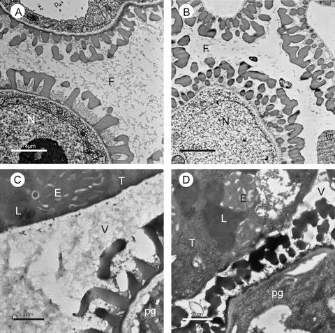
Figure 9.
Secretion Processes in Wild-Type and atgpat1-1 Tapeta.
Transmission electron micrographs of cross-sections of wild-type ([A] and [C]) and atgpat1-1 ([B] and [D]) anthers. At the ring-vacuolated microspore stage ([A] and [B]), a high concentration of fibrillar material is distributed uniformly throughout the wild-type locule (A), whereas much less material appears in the mutant locule (B). At the early bicellular stage ([C] and [D]), many osmophilic material–containing vesicles are secreted from the wild-type tapetum into the locule (C), whereas fewer vesicles are present in the mutant locule (D). E, elaioplast; F, fibrillar material; L, lipid body; N, nucleus; pg, pollen grain; T, tapetal cell; V, vesicle.

