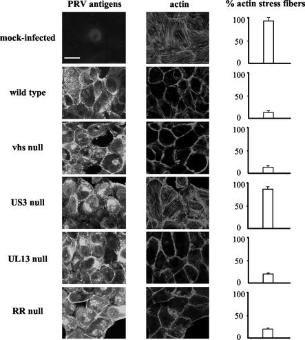
FIG. 1.
Actin architecture of SK-6 cells infected with different PRV strains. Confluent monolayers of SK-6 cells were mock infected or infected at an MOI of 10 with an NIA3 wild-type, NIA3 vhs null, NIA3 US3 null, NIA3 UL13 null, or NIA3 RR null strain. At 8 h p.i., cells were paraformaldehyde fixed, permeabilized, and stained with FITC-conjugated polyclonal PRV-specific antibodies and phalloidin-Texas Red to visualize PRV antigens (left panels) and actin filaments (middle panels), respectively. The right panels show the percentage of cells with intact actin stress fibers (200 cells were scored). Bar, 10 μm.

