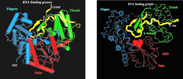
FIG. 3.
Overall structure of NS5B-570H. (Left) Ribbon representation of the polypeptide chain. The thumb domain is colored in green, the palm domain is in red, the fingers domain is in blue, and the C-terminal tail (residues 531 to 570) is in yellow. The putative RNA-binding groove is also indicated. (Right) Cα trace of NS5B-570H with the same color scheme as in the left panel. The C-terminal tail is highlighted in yellow ribbon. The catalytic-site -GDD- motif is shown as a red cpk ball model. The tip of the C-terminal tail reaches to the -GDD- pocket.

