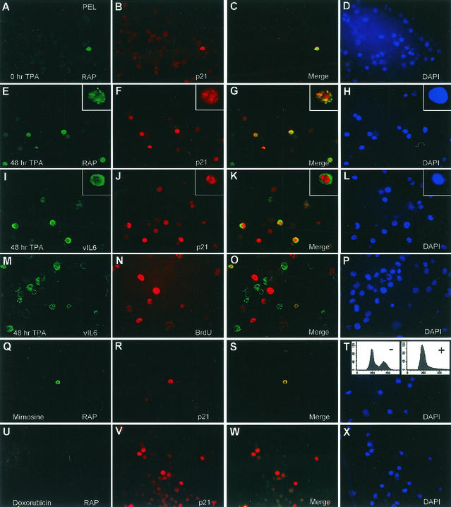
FIG. 9.
vIL-6 expression does not block RAP functions, and neither G1 cell cycle arrest nor p21 induction alone triggers the KSHV lytic cycle. (A to P) Double-label IFA experiments showing that TPA induction of vIL-6 during the lytic cycle in PEL cells does not interfere with RAP-mediated p21 induction and cell cycle arrest. (A to D) Uninduced BCBL-1 cells. Colocalized expression of both nuclear RAP (green) and p21 (red) proteins occurs in only a few spontaneously lytic cells. (E to L) TPA-induced BCBL-1 cells. Expression of p21 (red) occurs in a majority of the RAP-positive and vIL-6-positive cells (cytoplasmic, green). (M to P) TPA-induced BCBL-1 cells. S-phase BCBL-1 cells incorporating BrdU (red) do not colocalize with vIL-6-positive cells undergoing the lytic cycle. (Q to T) Uninduced BCBL-1 cells. Synchronization in G1 with mimosine does not trigger KSHV RAP expression. (Q) Detection of very few RAP-positive cells (green) in 30-h mimosine-treated BCBL1 cells. The inset FACS analysis shows the cell cycle profile of the same BCBL-1 cells before (−) and after (+) mimosine treatment. (R) Detection of p21-positive cells in the same field as in panel Q. (S) Merge of the two previous frames. (T) DAPI staining of the same field showing all cell nuclei. (U to X) Uninduced BCBL-1 cells. Doxorubicin-induced p21 expression does not trigger KSHV RAP expression in the same cells. (U) Detection of RAP-positive cells (green) after a 30-h doxorubicin treatment. (V) Detection of p21-positive cells in the same field. (W) Merge of the two previous frames. (X) DAPI stain showing all cell nuclei in the same field.

