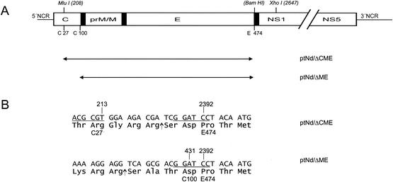
FIG. 1.
Structure of TBE virus replicons. (A) Schematic of the TBE virus genome (top) and the locations of the introduced deletions (below, double-headed arrows together with the corresponding plasmid designations). The open reading frame is represented by an open box, with only the structural protein coding region drawn to scale. Internal signal sequences are shown as black bars. Positions of cleavage sites of restriction enzymes used during cloning (nucleotide numbers refer to the wild-type genome of TBE virus) and amino acid residues flanking the deletions (numbering starts with the first amino acid residue of protein C or protein E) are indicated above and below the schematic, respectively. NCR, noncoding region. (B) Sequence details of the junction regions. Nucleotide and amino acid residues of the wild-type TBE virus sequence flanking the deletions are marked above and below the sequences, respectively. Recognition sites for restriction enzymes are underlined (MluI, ACG CGT; BamHI, GGA TCC). In the case of plasmid pTNdΔCME, 15 nucleotides coding for an artificial NS2B/3 protease cleavage site and creating the BamHI restriction site were introduced. The insertion of a T residue in plasmid pTNdΔME created a BamHI cleavage site (underlined) and maintained the authentic residue 100 (Asp) of protein C. Arrows depict the sites at which the amino acid sequences are presumed to be cleaved by the viral protease NS2B/3.

