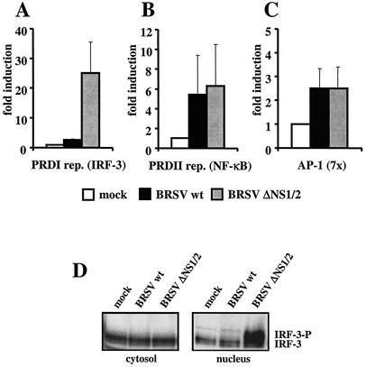
FIG. 6.
BRSV wt selectively blocks the activation of IRF-3. (A to C) HEK 293 cells were transfected with plasmids harboring the luciferase gene under the control of promoters containing either IRF-3 (A), NF-κB (B), or AP-1 (C) binding sites. Ten hours posttransfection, cells were infected with the indicated viruses at an MOI of 0.2. Fourteen hours postinfection cells were harvested and luciferase activity was determined. Relative light units are given as fold induction. Results show the mean values of four independent experiments with error bars showing standard deviations. (D) IRF-3 is not phosphorylated in BRSV wt-infected cells. HEK 293 cells were mock infected or infected with the indicated viruses at an MOI of 0.25. Ten hours postinfection, cells were harvested, and nuclear extracts were prepared as described in Materials and Methods and subjected to SDS-PAGE. Nonphosphorylated and phosphorylated forms of IRF-3 were detected with an anti-IRF-3 antibody (Dianova).

