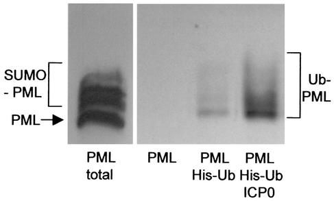
FIG. 1.
ICP0 induces the formation of ubiquitinated PML in transfected cells. HEp-2 cells were transfected with combinations of plasmids expressing epitope-tagged PML, polyhistidine-tagged ubiquitin with the lysine 48-to-arginine (K48R) mutation (His-Ub), and ICP0 (as indicated below the panels). The left panel shows a sample of a whole-cell extract of cells transfected with the PML expression plasmid alone (with the unmodified and SUMO-1-modified forms of PML indicated). The right panel shows a Western blot of ubiquitinated total cell proteins that had been purified by metal chelate affinity chromatography as described in Materials and Methods and probed for PML. PML was not detected in the left lane of this panel, which is a control with proteins purified from cells transfected with the PML plasmid in the absence of the tagged K48R ubiquitin plasmid. Inclusion of this plasmid results in the isolation of ubiquitin-modified PML bands (center lane). The quantity of these bands was greatly increased in the presence of coexpressed ICP0 (right lane).

