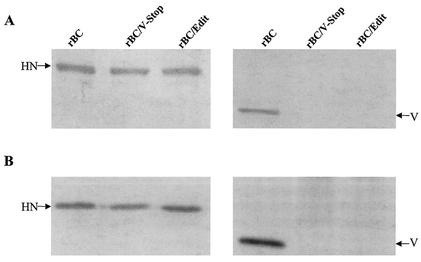
FIG. 2.
Western blot analysis performed with either purified viruses (A) or infected Vero cell extracts (B). Viral proteins were resolved in an SDS-12% polyacrylamide gel and transferred to polyvinylidene difluoride membranes in duplicate. The membranes were incubated with a cocktail of monoclonal HN antibodies or antipeptide serum specific for the carboxyl-terminal 18 amino acids of the V protein. The membrane was subsequently incubated with horseradish peroxidase-conjugated goat anti-mouse antibody or goat anti-rabbit IgG antibody, respectively. Reactive proteins were visualized in a visualization buffer.

