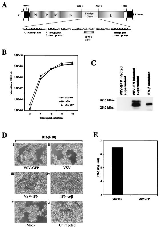
FIG. 1.
(A) Construction of VSV-IFNβ cDNA. Mouse IFN-β cDNA was inserted between the glycoprotein (G) and polymerase protein (L) genes of pVSV-XN2, a full-length cDNA clone of the Indiana serotype, as described in Materials and Methods. N, nucleocapsid protein gene; P, phosphoprotein gene; M, matrix protein gene. (B) One-step growth curves of recombinant viruses. BHK-21 cells were infected at an MOI of 10 PFU per cell with VSV-IFNβ, VSV-GFP, or rVSV not containing a foreign gene. The supernatants of infected cells were harvested at the indicated times, and viral titers were determined by a standard plaque assay. (C) Expression of IFN-β in VSV-IFNβ-infected cells. BHK-21 cells were infected with VSV-IFNβ or VSV-GFP at an MOI of 10 PFU per cell for 24 h. Supernatants (25 μl from 1 ml of 106 infected cells) were analyzed for IFN-β expression by immunoblotting using an anti-mouse IFN-β polyclonal antibody. Purified recombinant mouse IFN-β (UnitedStates Biological) (120 ng) was used as a positive control. (D) Biological assay of IFN-β expressed by VSV-IFN. BHK-21 cells were mock infected or infected with VSV-GFP, VSV-IFNβ, or rVSV at an MOI of 10 PFU per cell for 24 h. Supernatants (500 μl) were HI to remove residual virus and used to incubate B16(F10) cells for 24 h. Treated cells were then infected with wild-type VSV (Indiana strain) at an MOI of 0.1 for 24 h, and CPE was assessed under microscopy. (Di) B16(F10) cells treated with supernatants from VSV-GFP-infected BHK-21 cells; (Dii) B16(F10) cells treated with supernatants from rVSV-infected BHK-21 cells; (Diii) B16(F10) cells treated with supernatants from VSV-IFNβ-infected BHK-21 cells; (Div) B16(F10) cells treated with purified recombinant murine IFN-α/β (1,000 U/24 h); (Dv) B16(F10) cells treated with medium from uninfected BHK-21 cells; (Dvi) uninfected B16(F10) cells. (E) Quantitation of IFN-β expressed by VSV-IFNβ. The supernatant of BHK-21 cells infected with VSV-IFNβ or VSV-GFP at an MOI of 10 PFU per cell for 24 h was serially diluted and used to incubate C57BL/6 primary cells for 24 h. The cells were infected with wild-type VSV (Indiana strain) at an MOI of 0.01 PFU per cell for 24 h, and CPE was assessed by microscopy. Biological activity data are presented as units per milliliter and represent the means from two experiments.

