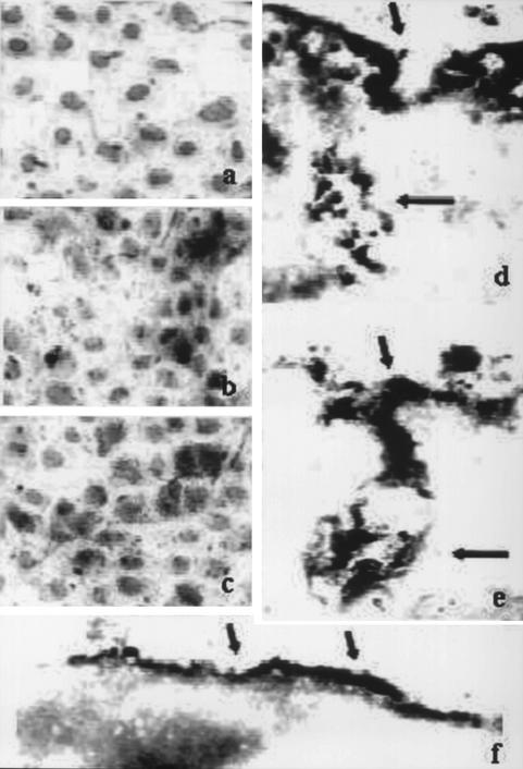
FIG. 2.
Detection of human IgG and FcRn receptor by immunocytochemistry and light microscopy (a to c; magnification, ×65,000) or by IEM (d to f; magnification, ×72,000). (a, b, d, and e) Detection of IgG in HuH-7 cells cultured in the absence of human IgG (a), in the presence of 1.0 mg of polyclonal anti-HBs IgG/ml (b and e), or in medium containing human AB serum (d). (c and f) Detection of human FcRn. Hematoxylin staining of nuclei (light grey) and peroxidase staining (black) are shown. The arrows (the short arrows indicate the cellular membrane; the long arrows indicate membranous invaginations) indicate the positive signals for IgG or FcRn.

