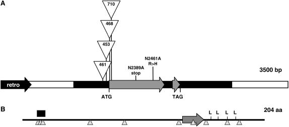
Figure 2.
dlf1 genomic organization and protein structure. A, Diagram of the genomic organization of dlf1. The white line indicates the unique 5′ and 3′ sequences flanking the dlf1 coding sequence (gray arrows). The black arrow (retro) denotes the closest 5′ retroelement. The thick black line indicates, from left to right, the 5′-UTR, the single intron, and the 3′-UTR. The positions of the start (ATG) and stop (TAG) codons are noted. The insertion sites for four of the dlf1-mu alleles are indicated as are the position of the two EMS alleles. B, Organization of DLF1 protein motifs. The basic domain is indicated by the large gray arrow, the Leu zipper by the four Leu, a Ser-rich domain by the black box, and the position of putative Ser/Thr phosphorylation sites by upward-pointing triangles.

