Figure 1.
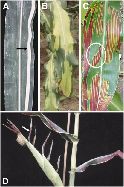
Clonal and nonclonal sectors in maize leaves. A, Arrow indicates clonal sector of white tissue illustrating longitudinal arrangement of cell files in a maize leaf. B, Variegated tdy1 mutant leaf with yellow and green nonclonal sectors. C, tdy1 leaf in anthocyanin-accumulating genetic background showing that yellow sectors accumulate red anthocyanins. White circle shows a regional island sector located adjacent to tissue of the opposite sector type. D, tdy1 red sectors occur only in leaf blade tissues; leaf sheaths and husk leaf sheaths do not develop sectors. White arrow indicates a tdy1 sector in leaf blade tissue at the tip of an ear husk leaf.

