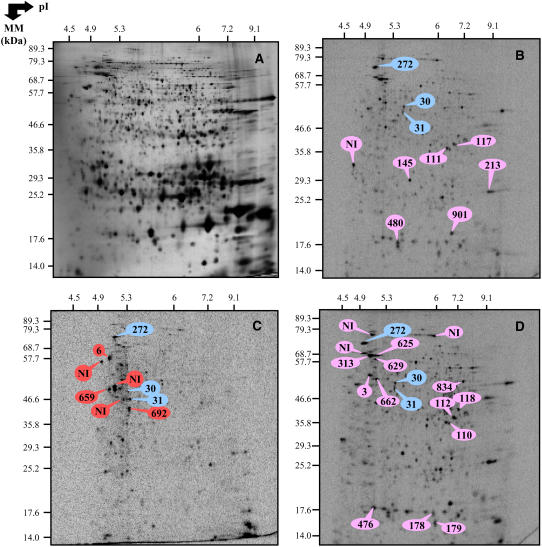
Figure 6.
2D patterns of de novo protein synthesis in D and ND seeds imbibed for 1 d on basal medium and in ND seeds imbibed for 1 d in the presence of 30 μm ABA. Radiolabeling of proteins was effected by adding [35S]-Met in the germination assays. Total soluble proteins were extracted from seed samples shown in Figure 5A, submitted to 2D gel electrophoresis and the radiolabeled proteins revealed by Phosphorimager analysis as described under “Materials and Methods.” A, Silver-stained 2D gel of total soluble proteins from the ND seeds imbibed for 1 d in basal medium. B, Autoradiogram of the 2 D gel for proteins extracted from the ND seeds imbibed in basal medium for 1 d. C, Autoradiogram of the 2 D gel for proteins extracted from the D seeds imbibed in basal medium for 1 d. D, Autoradiogram of the 2 D gel for proteins extracted from the ND seeds imbibed for 1 d in the presence of 30 μm ABA. The labeled protein spots were identified either by MS or by comparison with Arabidopsis seed protein reference maps from Ler (Rajjou et al., 2006a; http://seed.proteome.free.fr; see Supplemental Tables S3 and S5). They are listed in Supplemental Table S4. The proteins labeled in blue color correspond to proteins synthesized de novo in the D and ND seeds imbibed in basal medium for 1 d and in the ND seeds imbibed for 1 d in the presence of 30 μm ABA (no. 30, enolase; no. 31, Xyl isomerase; no. 272, MUT/nudix family protein). The proteins labeled in red color correspond to proteins synthesized de novo in the D seeds imbibed for 1 d in basal medium (no. 6, vacuolar ATP synthase α-chain; no. 692, hydrolase alpha β-chain; no. 659, 26S protease regulatory family). The proteins labeled in pink color correspond to proteins synthesized de novo in the ND seeds imbibed for 1 d in basal medium or in the presence of 30 μm ABA.

