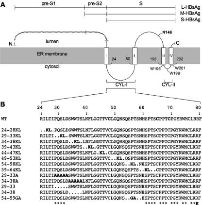
FIG. 1.
Schematic representations of envelope protein mutants. (A) L-, M-, and S-HBsAg proteins are depicted by horizontal lines. The topology of the envelope protein S domains at the ER membrane is represented. Open boxes represent transmembrane regions in the envelope proteins S domain, and the shaded area corresponds to the ER membrane. Experimentally defined (I and II) or putative (III and IV) transmembrane signals are indicated. (B) CYL-I sequences of the envelope protein mutants are indicated. Mutants are designated by the positions of the first and last deleted amino acids followed by the one-letter codes of the inserted residues. Asterisks indicate the positions of amino acids that were substituted with alanine in mutants presenting single amino acid substitutions. The R79K mutation (K) is indicated.

