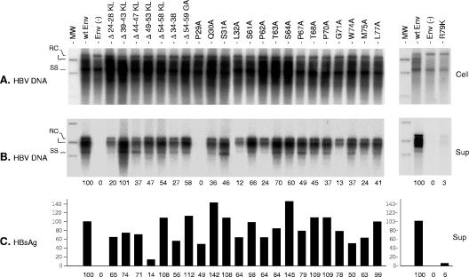
FIG. 3.
Effects of envelope protein CYL-I mutations on secretion of HBV virions. (A) Southern blot analysis of intracellular HBV DNA. Total DNA from HuH-7 cells (Cell) cotransfected with pCIHBenv(−) and pT7HB2.7 or derivatives was extracted at day 11, separated on a 1% agarose gel, transferred to a nylon membrane, and probed with a 32P-RNA probe specific for negative-strand HBV DNA. Signals are from 105 cells. Molecular weight markers (MW) include 10 pg of cloned linear (L) HBV DNA, corresponding to 3 × 10⁶ ge. (B) Culture media collected at days 3, 6, 9, and 11 posttransfection (Sup) were pooled and subjected to immunoprecipitation using an anti-preS1 antibody. Precipitates were disrupted, and DNA was extracted and analyzed by Southern blot hybridization as described above. Signals are from 1 ml of culture medium. For a positive control (wt Env), the 100% values correspond to 200 pg/ml of HBV DNA (left panel) or 300 pg/ml (right panel), approximately 6 × 10⁷ or 9 × 10⁷ ge/ml, respectively. (C) Histogram showing the relative amounts of secreted HBV envelope proteins measured with an anti-HBsAg ELISA (DiaSorin). For a positive control (wt Env), the 100% value corresponds to approximately 400 ng/ml of HBsAg (left panel) or 150 ng/ml (right panel), equivalent to approximately 1011 or 3.7 × 10¹⁰ SVPs, respectively. RC, relax circular; L, linear; SS, single stranded; Env(−), envelope protein negative; wt SML, wt S-, M-, and L-HBsAg. Numbers below each panel are percentages of the wt value.

