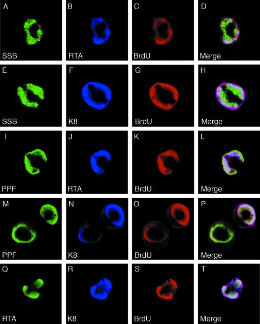
FIG. 10.
Visualization and characterization of VRC in TPA-induced BCBL-1 cells and localization of RTA and K8 in VRC. BCBL-1 cells were treated with TPA for 48 h and pulse-labeled with BrdU for 60 min. The cells were subjected to triple-label IFA using mouse monoclonal anti-RTA or anti-K8 antibody (Cy5), rabbit polyclonal anti-SSB or anti-PPF antibody (FITC), and sheep anti-BrdU antibody (Texas Red). The triple-label IFA shows that RTA and K8 are colocalized with core replication machinery proteins (SSB and PPF) as well as newly synthesized DNA (BrdU incorporated) in VRC in TPA-induced BCBL-1 cells.

