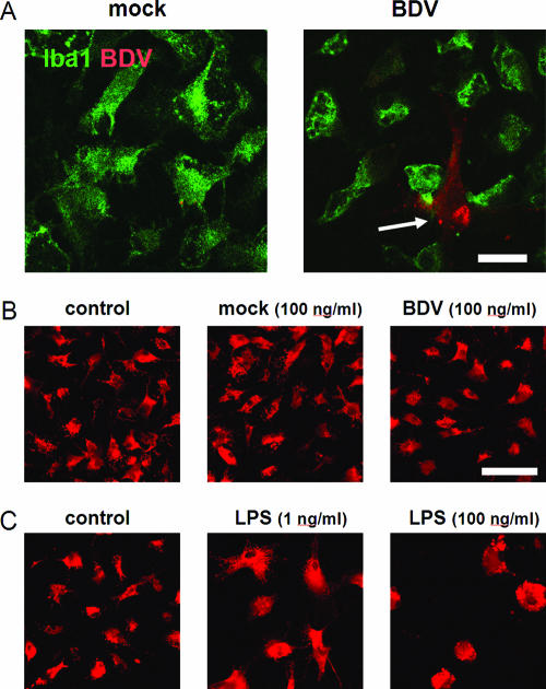
FIG. 1.
Cultured microglia are neither infected nor activated by purified BDV. (A) Microglia were exposed to the purified mock stock (left) or BDV stocks at a MOI of 0.1 (right) for 5 days. Images show immunostaining for a microglial marker, Iba1 (green), and BDV N (red). Note the lack of colocalization of the Iba1- and BDV-positive cells. The arrow points to an infected nonmicroglial cell occasionally present in the microglia cultures. (B) Exposure of microglia to the mock- or BDV-infected stocks did not resulted in change of the cell morphology or the ED1 expression. (C) LPS treatment induced a dose-dependent change in the cell shape. In panels B and C immunostaining for activated the macrophage/microglia activation marker ED1 (red) is shown. The data shown represent the 5-day cultures. Scale bar, 40 μm.

