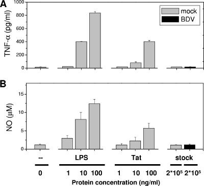
FIG. 2.
LPS and HIV-Tat but not purified BDV activate microglia to produce TNF-α and NO. Microglia cultures (1-day in vitro) were exposed to cell-free purified BDV or mock stock for 2 days in the presence or absence of LPS (1, 10, 100 ng/ml) or HIV-Tat protein (1, 10, 100 ng/ml). The concentrations of the proinflammatory molecules TNF-α (A) and NO (B) released by the microglia were measured following incubation with LPS (1, 10, 100 ng/ml), Tat (1, 10, 100 ng/ml), the cell-free purified BDV (200 μg/ml which is equivalent to a MOI of 0.1) or mock stocks (200 μg/ml). Untreated cultures (zero) served as the negative control. The results of a representative experiment are shown, and data are the means ± standard error of the means for two wells of one 24-well plate. Duplicate experiments yielded similar results. Note the elevated secretion of NO and TNF-α following treatments with LPS or the Tat protein but not following incubation with the purified virus or BDV-infected neuronal cells.

