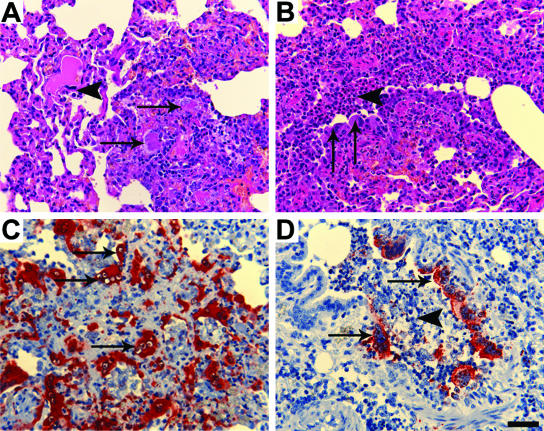
FIG. 2.
Histopathology and immunohistopathology associated with NiV infection in cats. (A) Necrotizing alveolitis (arrows) in NiV-infected cat and endothelial syncytial cell (arrowhead) (HE). (B) Broncho-alveolitis in NiV-infected cat, with bronchiolar epithelial syncytial cells (arrows) and intraluminal debris (arrowhead) (HE). (C) Positive staining for NiV antigen in regions of alveolitis, including endothelial syncytial cells (arrows) (anti-NiV polyclonal antibody). (D) Positive staining for NiV antigen in bronchiolar epithelial cells (arrows) and intraluminal debris (arrowhead) (anti-NiV polyclonal antibody). Scale bar for all images = 50 μm.

