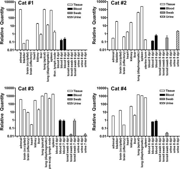
FIG. 3.
NiV genome in cats detected by TaqMan PCR. Normalized, relative NiV genome levels in samples collecting during NiV infection in cats and at necropsy. TaqMan PCR CT values were determined in triplicate for the NiV genome and normalized by dividing this value by the 18S rRNA CT values for each sample. The relative NiV genome was determined by linear regression of NiV cDNA standard curves for each assay. Values are expressed as the average of all replicates. The adrenal gland, liver, lung, spleen, and lymph nodes consistently displayed the highest relative NiV genome levels, while the brain and heart frequently revealed the lowest. The genome was detectable in the blood in all cats 1 day prior to euthanasia but only detectable in the urine from cats infected with 5,000 TCID50 of NiV.

