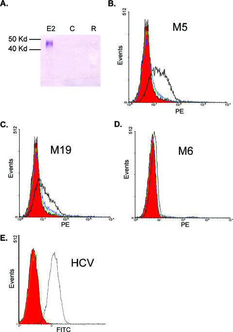
FIG. 1.
Purification of GBV-C E2 protein and development of a cell binding assay. (A) Immunoblot assay of culture supernatant from CHO cells expressing E2 (lane E2), CHO cells without E2 (lane C), or Roche cell lysates containing E2 (lane R). E2 detected with anti-E2 MAb M6. Kd, kilodaltons. (B to D) GBV-C E2 protein (10 μg/ml) was incubated with MOLT-4 cells, and cell-bound E2 was detected by GBV-C anti-E2 MAbs M5 (B), M19 (C), and M6 (D) (all with open black lines). Controls (isotype control antibody and anti-HCV E2 MAb, respectively) are shown in red and green. Cells incubated with HCV E2 (10 μg/ml) and detected with M5 (B), M19 (C), and M6 (D) are shown in blue. Panel E demonstrates HCV E2 binding to MOLT-4 cells including isotype control antibody (red), HCV MAb (black), or GBV-C M5 (green).

