Figure 9.
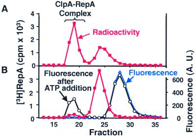
Inhibition of RepA-ClpA complex formation by unfolded GFP. (A) ClpA (100 pmol) was incubated (in 100 μl) of buffer A containing 2 mM ATP[γS] for 5 min at 24°C. Then 100 pmol of [3H]RepA (1,170 cpm/pmol) was added and the reaction mixture was incubated for 10 min at 25°C. The mixture was applied to a 0.7 × 15 cm column of Sephacryl S200 HR as described in the legend of Fig. 3. Radioactivity was measured in aliquots of the fractions (red squares). (B) ClpA (100 pmol) was incubated as in A with the exception that 300 pmol of acid-denatured GFP was added to the initial reaction mixture. After incubation with [3H]RepA as in A, the sample was analyzed by gel filtration as above. Radioactivity was measured (red squares). Initial fluorescence of the fractions was measured (open blue triangles). Then, 5 mM ATP and an ATP-regenerating system were added and fluorescence was measured after 15 min at 24°C (open black circles; A. U., arbitrary units).

