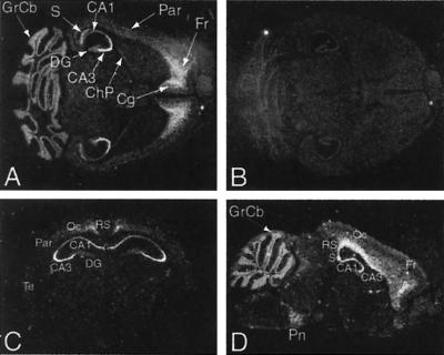
Figure 8.
In situ hybridization of adult mouse brain. Darkfield autoradiographs of representative horizontal (A and B), coronal (C), and parasagittal (D) sections hybridized with Sebox antisense (A, C, and D) or sense (B) [33P]-labeled RNA probes. (A) Expression of Sebox RNA was detected in the cerebral cortex, CA areas of the hippocampus, subiculum, choroid plexus, and granular layer of the cerebellum. Within the hippocampus, intense labeling is present in the pyramidal cells of the CA3 region (A and C), whereas no Sebox RNA was detected in the granule cells of the dentate gyrus. In the cerebral cortex, Sebox is expressed at high levels in the frontal and cingulate cortex (A) and, more posteriorly, in the retrosplenial and occipital cortex (C and D). A lower level of Sebox mRNA was detected in the parietal and temporal cortex (A and C). In D, Sebox expression is shown in a continuous pattern that covers the CA regions of the hippocampus, the subiculum, and a deep layer of the cerebral cortex (retrosplenial, occipital, and frontal cortex). The pontine nuclei also are labeled (D). Abbreviations are as follows: CA1–3, CA regions of the hippocampus; ChP, choroid plexus; Cg, cingulate cortex; DG, dentate gyrus; Fr, frontal cortex; GrCb, granular layer of the cerebellum; Oc, occipital cortex; Par, parietal cortex; Pn, pontine nuclei; RS, retrosplenial cortex; S, subiculum; Te, temporal cortex.

