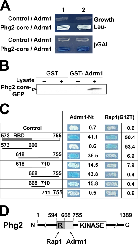
Figure 6.
A specific domain of Phg2 interacts with the Adrm1 protein. (A) Two distinct clones (1 and 2) expressing the indicated proteins are shown. Growth in Leu- medium (top panel) as well as production of β-galactosidase (blue color, lower panel) indicated that the core region of Phg2 interacted with the Adrm1 protein in a yeast two-hybrid assay. (B) A GST-Adrm1 fusion protein was immobilized on glutathione beads and mixed with a lysate of Dictyostelium cells overexpressing the Phg2 core region tagged with GFP. Binding of the GFP fusion protein to GST-Adrm1 beads was detected with an anti-GFP antibody. (C) A collection of mutants of the Phg2 core region was constructed, and their interaction with Adrm1 and Rap1 was tested in a two-hybrid assay. The production of β-galactosidase was visualized on plates. It was also evaluated more precisely in a liquid assay and is indicated for each construct (arbitrary units). (D) Schematic representation of Phg2. The core region contains two distinct regions: the RBD (R) interacts with Rap1, whereas the adjacent region interacts with Adrm1.

