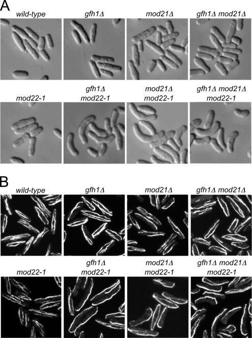
Figure 1.
Cell shape and microtubule distribution in gfh1Δ, mod21Δ, and mod22-1 single and double mutants. (A) Cell shape in strains of the indicated genotypes after growth to stationary phase on solid media, replica-plating to fresh media, and subsequent growth for 3 h. (B) Anti-tubulin immunofluorescence of asynchronous, exponentially growing cells of the indicated genotypes.

