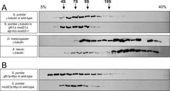
Figure 12.
Fission yeast γ-tubulin is mostly present in a small complex on sucrose gradients. Western blots of cell extracts after sucrose gradient sedimentation. (A) Untagged γ-tubulin in extracts from wild-type and gfh1Δ mod21Δ alp16Δ mod22-1 fission yeast as well as from Drosophila embryos and Xenopus eggs. (B) Myc-tagged gfh1p, and, independently, Myc-tagged mod21p, in extracts from wild-type fission yeast. The top of the gradient is at the left, and the positions of S-value standards are indicated above the top panel. The gap in staining near the bottom of the Drosophila extract gradient is due to a loading error.

