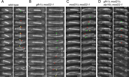
Figure 9.
Microtubule behavior at the end of mitosis. Stills from movies of wild-type (A) and mutant (B–D) cells toward the end of mitosis. Time indicates minutes and seconds relative to the first time point shown; unless indicated, the time between successive frames is 30 s. Note that astral microtubules tend to be short-lived. Arrow in B indicates rare release of an astral microtubule from the spindle pole body. Note also that microtubules are sporadically “fired” from the equatorial MTOC in the center of the cell before formation of a well-formed postanaphase array. Colored dots indicate the presumed minus ends of these microtubules, with a single color for each such microtubule. In some cases, these microtubules seem to translocate away from the nucleation site, both in wild-type and mutant cells. All cells shown express both GFP-atb2p and endogenous atb2p.

