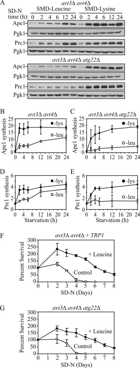
Figure 7.
Protein synthesis and survival dependent on autophagic amino acids was reduced in avt3Δ avt4Δ mutant cells, and the defect was exacerbated by the atg22Δ mutation. (A) avt3Δ avt4Δ (ZFY20) and avt3Δ avt4Δ atg22Δ (ZFY22) cells were grown and analyzed for up-regulated protein synthesis as described in the legend to Figure 6. (B–E) Quantification of immunoblots from SMD-leucine (open symbols) or SMD-lysine (closed symbols) in the avt3Δ avt4Δ (square), or avt3Δ avt4Δ atg22Δ (circle) cells. Band intensities were quantified as described in the legend to Figure 6. The loss of leucine effluxers blocked synthesis of Ape1 and Prc1 during leucine depletion. The avt3Δ avt4Δ strain harboring pRS414 (TRP1) (F) and avt3Δ avt4Δ atg22Δ (G) cells were grown in SMD to mid-log phase and incubated in SD-N or SD-N containing 100 μg/ml leucine. At the indicated days, viability was determined as described in the legend to Figure 4. The error bars indicate the SD of three independent experiments. The reduced viability of the avt3Δ avt4Δ, and avt3Δ avt4Δ atg22Δ mutant cells was partially rescued by addition of leucine.

