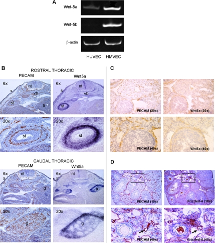
Figure 1.
Wnt5a and Frizzled-4 expression in vascular cells. (A) Wnt5a and Wnt-5b RT-PCR products from human primary endothelial cells, HUVEC and HMVEC, normalized to β-actin transcript levels. (B) In situ hybridization in mouse embryonic tissue at E13.5 for PECAM and Wnt5a. nt, neural tube; li, liver; st, stomach; vb, vertebral body; g, gonad; s, skin. (C) Immunohistochemistry with PECAM (left) and in situ hybridization with Wnt5a mRNA probe (right) in mouse embryonic tissues at E13.5. Brown color represents PECAM protein (left panels), and the nuclei are counterstained in blue. Wnt5a signal is seen in blue color on the in situ (right) panels. Both immunohistochemistry and in situ hybridizations were performed using adjacent serial sections. (D) Adult mouse ovaries were immunostained for either PECAM or Frizzled-4 to compare expression. Top, insets indicate the area shown at higher magnification in the bottom panels; bottom, arrows indicate blood vessels.

