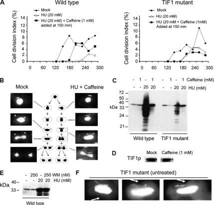
Figure 6.
Identification of an intra-S-phase checkpoint defect in Tif1p-deficient T. thermophila. (A) Wild-type (CU428) and tif1-1::neo/ TIF1 mutant (TXh48) strains were grown to saturation, starved, and then released into drug-free media (filled diamonds) or media containing 20 mM HU (open triangles and filled squares). Caffeine (1 mM) was added before the onset of macronuclear S phase (T = 150 min) (filled squares), and cell division was monitored by light microscopy. The cell division index corresponds to the percentage of cells with a cytokinetic furrow. (B) DAPI analysis of micro- and macronuclear division in mock and HU + caffeine-treated wild-type cells (same treatment as in A). (C) Rad51p Western blot analysis in synchronous wild-type and TIF1 mutant cultures, 5 h after refeeding with media containing HU, caffeine, or both. (D) Tif1p Western blot analysis of untreated and caffeine-treated wild-type strain (CU428; 1 mM caffeine for 4 h). (E) Rad51p Western blot analysis in synchronous wild-type cultures, 5 h after refeeding with media containing WM, HU, or both. (F) DAPI analysis documenting aberrant micro- and macronuclear division in tif1-1::neo mutant cells (TXh48) grown in normal culture media (no HU added). Arrow: cytokinetic furrow.

