Figure 3.
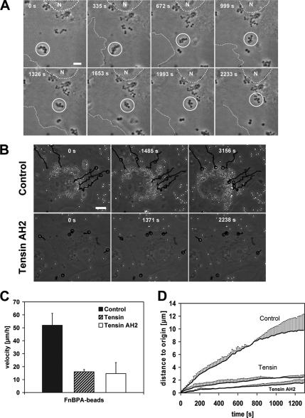
FnBPA-mediated bacterial movements are controlled by tensin. (A) Movement of an FnBPA-S. carnosus cluster on HUVECs. Cells were infected with FnBPA-S. carnosus and subjected to live cell imaging (details in Materials and Methods). The depicted phase contrast images were taken from a movie at the indicated time points (Supplemental Movie 1). Circle highlights the position of the moving bacterial cluster. Dotted line outlines the nucleus (N) and broken line the cell margin. Bar, 5 μm. (B) Effect of tensin AH2 overexpression on FnBPA-bead movements. HUVECs expressing GFP (control) or GFP-tensin AH2 were infected with FnBPA-beads and subjected to live cell imaging. The depicted phase-contrast images were taken from the movies at the indicated time points (Supplemental Movies 2 and 3). Tracks of selected FnBPA-beads are indicated by lines. Circle points out the actual position of the beads on their track. Bar, 10 μm. (C) Velocity of FnBPA-beads in HUVECs overexpressing tensin or tensin AH2. GFP-, GFP-tensin-, or GFP-tensin AH2-expressing HUVECs were infected with FnBPA-beads and subjected to live cell imaging. Bead movements were tracked and velocities were calculated. Each bar represents mean ± SD from nine (GFP-tensin), 21 (GFP-tensin AH2), or 19 (GFP/control) values obtained in two to four different experiments. (D) Same experiment as in C, but the distances that beads covered relative to the starting point (0 s) are depicted.

