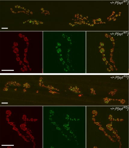
Figure 2.
GluRIII and nc82 antibody staining was unchanged in polylysine motif mutants. Representative 40 and 63× images of neuromuscular junctions from transgenic controls (−/−;P[sytWT], top) and polylysine motif mutants (−/−;P[sytKQ], bottom). Junctions were stained with an anti-GluRIII antibody (red) to show postsynaptic receptors, and they were stained with mAb nc82 (green) to show active zones. Bar, 10 μm.

