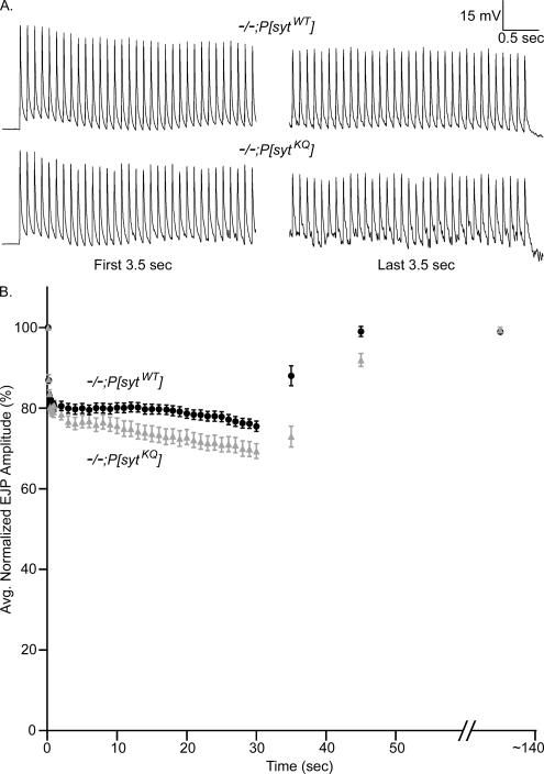
Figure 7.
Polylysine motif mutants recover more slowly from synaptic depression than transgenic controls. Synaptic depression was elicited by stimulating fibers at 10 Hz for 30 s in 5 mM extracellular Ca2+. Recovery was monitored by recording the EJP amplitude of a single shock either 5 or 15 s after the 10-Hz stimulation stopped. Each fiber was also stimulated at an extended time point (∼140 s) to verify full recovery. (A) Representative traces from polylysine motif mutants (−/−;P[sytKQ]) and transgenic controls (−/−;P[sytWT]) showing the first and last 3.5 s of stimulation at 10 Hz for 30 s in saline containing 5 mM Ca2+. (B) EJP amplitudes were normalized to the amplitude of the first shock. After the first nine points, the points plotted during the 10-Hz stimulation are averages of 10 EJPs. Transgenic controls: black circles, n = 10 fibers for 5-s recovery and n = 14 fibers for 15-s recovery. Polylysine motif mutants: gray triangles, n = 10 fibers for 5-s recovery and n = 14 fibers for 15-s recovery. Error bars are SEM.

