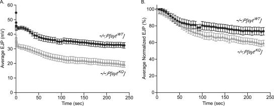
Figure 8.
Polylysine motif mutants maintain significant levels of evoked release during prolonged high-frequency stimulation. (A) Average EJP amplitudes evoked during 4 min of 10-Hz stimulation in saline containing 5 mM Ca2+. Except for the first nine points and the last point, the EJP amplitudes were binned at 5-s intervals, and the average of the bin is plotted. The first nine points are not binned. The last point is the average EJP amplitude of a 3-s bin. Transgenic controls: black circles, n = 8 fibers. Polylysine motif mutants: gray triangles, n = 10 fibers. (B) Same EJP amplitudes as in A but binned at 5-s intervals and normalized to the first 5-s bin.

