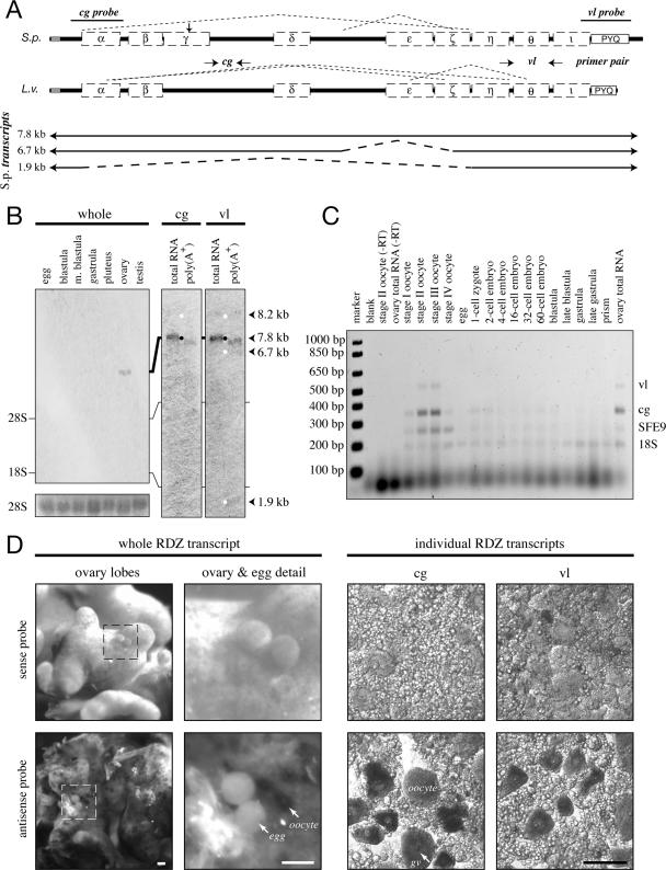
Figure 1.
Rendezvin transcripts are exclusive to oocytes. (A, top) Schematic identifying splicing organization predicted by RT-PCR fragments in Figure 2 (dashed lines), probe locations (S. purpuratus), and primer pairs (L. variegatus) relative to the predicted open reading frames, including motifs (see Supplemental Figure 1). cg, cortical granule; vl, vitelline layer. (A, bottom) Diagram of S. purpuratus coding strand RNA identified by sequenced RT-PCR products, listed with likely size of each transcript based on RNA blot pattern. (B) RNA blots probing S. purpuratus total RNA (10 μg) from different developmental stages and gonads, or S. purpuratus ovary total RNA (2.5 μg) and oocyte-enriched poly-A+ RNA (1 μg). The developmental blot (left) was probed with an equal mixture of antisense RNA probes. Duplicate RNA blots (right) were probed with either rdz probe and then overexposed to identify all major transcripts (white dots). Approximate size of each identified transcript is also shown, calculated based on migration distance (our unpublished data). rRNA bands are indicated. Ethidium bromide staining for the 28S rRNA bands is shown as loading controls for the developmental blot. (C) Ethidium bromide image of multiplex reverse transcriptase PCR amplifications from two-cell/embryo equivalents of whole L. variegatus cells or embryos, or 250 ng of ovary total RNA. Primer pairs for the vitelline layer or cortical granule transcript of rendezvin, the cortical granule protein SFE9, and the 18S rRNA (Supplemental Table 3) were used to amplify specific products from total lysates of each stage indicated. Oocyte staging was based on the relative nuclear-to-cytoplasmic ratio, as described previously (Wong et al., 2004). Controls include no template (“blank”) and reactions without reverse transcriptase (“-RT”). (D) In situ hybridizations of rendezvin on S. purpuratus ovary and eggs. Left set shows intact ovary lobes and ovulated eggs, including a magnified image. Right set shows tissue smears of ovary hybridized with either probe (see A). Both sense (top images) and antisense (bottom images) are shown. Oocytes (oo), their germinal vesicles (gv), and eggs are labeled. Bars, 100 μm.

