Figure 4.
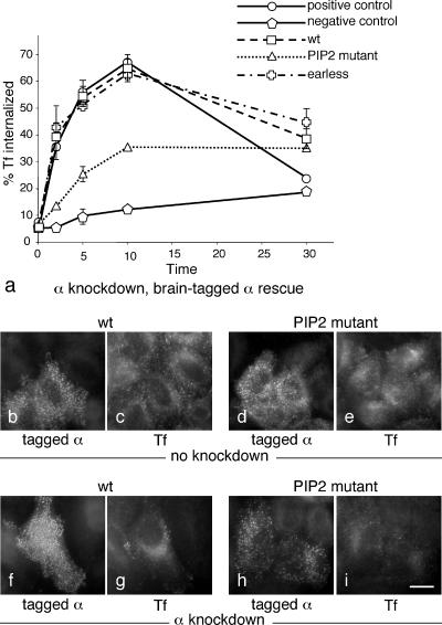
Receptor-mediated endocytosis of transferrin in cells expressing the brain-tagged α constructs. (a) Cells were allowed to bind 125I-labeled transferrin at 4°C and then shifted to 37°C for the indicated length of time, after which the medium was harvested, ligand remaining on the cell surface was released with an acid wash, the cells were solubilized with NaOH, and all three fractions were quantified and expressed as % total counts. The graph shows the percentage of the total counts recovered in the NaOH extract (i.e., internalized transferrin). The cells used for the positive control were neither transfected nor treated with siRNA; the cells used for the negative control were treated with siRNA only. All other cells were stable cell lines expressing brain-tagged α constructs, which were depleted of endogenous α using siRNA. The wild-type and earless constructs fully rescue the knockdown phenotype; the PtdIns(4,5)P2 binding mutant gives partial rescue. (b–i) Cells were transiently with either the wild-type α construct (b, c, f, and g) or the PtdIns(4,5)P2 binding mutant (d, e, h, and i), either with (f–i) or without (b–e) concurrently knocking down endogenous α. Alexa Fluor 594–conjugated transferrin was added to the cells for 10 min at 37°C. Neither construct has a dominant negative effect, and they both appear to give at least partial rescue, consistent with the biochemical data on stably transfected cells shown in a. Scale bar, 20 μm.

