Figure 2.
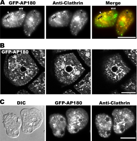
Localization of clathrin and AP180. (A) AP180 and clathrin colocalize. Cells expressing GFP-AP180 were fixed and stained with an anti-clathrin light-chain antibody detected with a Texas Red–conjugated secondary antibody. The majority of AP180 punctae at the plasma membrane colocalized with clathrin (black arrows) although some AP180 punctae lacked clathrin (white arrows). Merge shows GFP-AP180 in green and clathrin in red. Scale bar, 10 μm. (B) Z-series of cells expressing GFP-AP180 fixed and imaged by confocal microscopy. GFP-AP180 punctae decorated the contractile vacuole bladder (arrow, left and middle panels) as well as the tubules radiating from it (arrows, right panel). The two bright signals adjacent to the central bladder represent two nuclei. Each panel is 0.8 μm apart. Scale bar, 10 μm. (C) Cells expressing GFP-AP180 were fixed and stained with an anti-clathrin light-chain antibody followed by Texas Red–conjugated secondary antibody. Punctae of GFP-AP180 and clathrin outlined the contractile vacuole (arrow). Scale bar, 10 μm.

