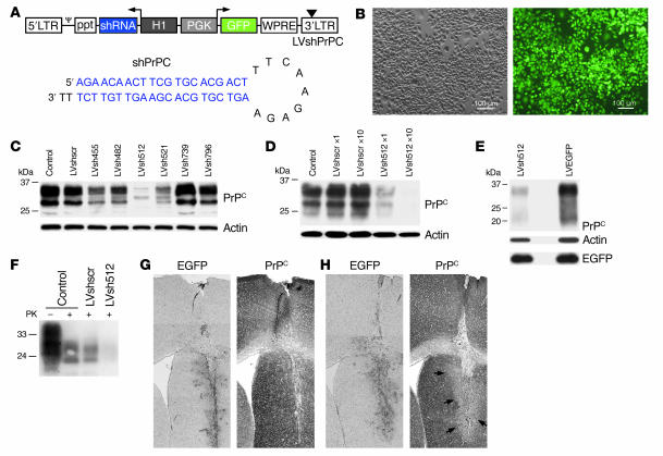
Figure 1. Targeting of PrPC by lentivector-mediated RNAi.
(A) The HIV-based lentivector (LVshPrPC, top) used carries an H1 promoter–driven shRNA cassette and a PGK-EGFP expression cassette. Ψ, packaging signal; ppt, polypurine tract; WPRE, woodchuck hepatitis virus posttranscriptional regulatory element; triangle, self-inactivating mutation; arrows, direction of transcription. The lentivector-encoded anti-PrPC shRNA (shPrPC, bottom) consists of 19- or 21-bp stems separated by a loop. (B) Representative bright-field (left) and fluorescence microscope (right) images of N2a cells 72 hours after transduction with LVshPrPC. (C) LVshPrPC-induced knock down of PrPC in N2a cells. Control, uninfected cells. (D) PrPC expression in N2a cells infected with a low (×1) and high (×10) concentration of LVsh512 and LVshscr. (E) Analysis of PrPC expression in granule cells 72 hours after infection with LVsh512 or a control vector carrying only the EGFP expression cassette (LVEGFP). Western blots (C–E) were probed with the indicated antibodies. (F) Effect of LVsh512 on the accumulation of protease K–resistant (PK-resistant) PrPSc in ScN2a cells. Western blot analysis 72 hours after transduction with the indicated lentivectors. (G and H) Intracranial injection of LVshscr (G) and LVsh512 (H) in tga20 mice overexpressing PrPC. Analysis of EGFP (left) and PrPC (right) expression 3 weeks after injection. Arrows indicate the lentivector-transduced area.

