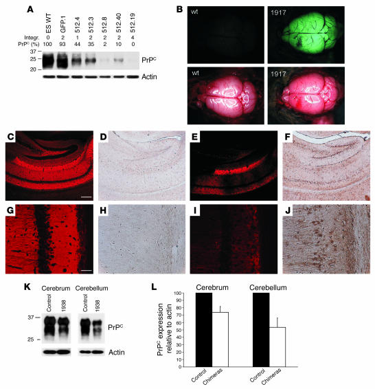
Figure 2. Silencing of PrPC in chimeric mice derived from LVsh512-infected ES cells.
(A) Western blot analysis of ES cell clones infected with LVsh512 or LVEGFP. The number of integrants (Integr.) and levels of PrPC expression (PrPC) are given above the blot. (B) Fluorescence imaging of freshly isolated brains from a control mouse (WT, left) and a transgenic animal (no. 1917, right). Shown are the fluorescence (top) and the bright-field (bottom) images. (C–J) Immunohistochemical analysis of PrPC and EGFP expression in sections of hippocampus from a transgenic (no. 1917) and an age-matched WT animal. (C and G) Analysis of PrPC expression in WT hippocampus. (E and I) Staining for PrPC revealed reduced expression of PrPC in the chimeric hippocampus as compared with the WT. (D and H) Expression of EGFP in the WT mouse. (F and J) Staining for EGFP in the chimeric hippocampus, indicating the presence of the LVsh512 provirus. Higher magnifications are shown in G and H for the WT and I and J for the chimeric mouse. Scale bars: 200 μm in C–F and 50 μm in G–J. (K and L) Western blot analysis of PrPC expression in the cerebrum and cerebellum of chimeric animals.

