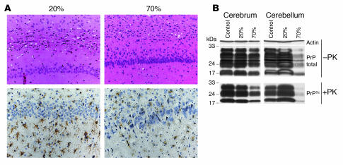
Figure 4. Analysis of PrPSc accumulation and neuropathological changes in prion-inoculated chimeric siblings.
(A) Histological analysis of a scrapie-infected chimeric mouse (20%) that died at 172 dpi and a 70% chimera that was sacrificed at the same time. Analysis of spongiform changes (top) and gliosis (bottom). Coronal sections of the posterior cerebrum at the hippocampal level of the 20% (left) and 70% (right) chimeric siblings are shown. Top: H&E staining; bottom: glial fibrillary acidic protein (GFAP) staining (brown). (B) Western blots of samples from the same chimeric mice as shown in A and a wild-type control. Levels of PrPSc (+PK) and total PrP (PrPtotal, –PK) in 2 different brain regions: cerebrum including hippocampus (left) and cerebellum (right).

